
Bone health after fracture
Bone Health after Fracture
A fracture can be a broken bone that needs treatment. It can also be a signal that the skeleton deserves attention. This pathway explains what to ask after a wrist, hip, shoulder, spine compression, or other low-energy fracture so the fracture care and bone-health care move together.
Visual explainer
A fracture can be the first visible clue that bone strength deserves attention.
These are examples of real fragility-type fracture radiographs from open-access clinical articles. They are not here for self-diagnosis; they show why fracture follow-up should include the skeleton, not only the broken bone.
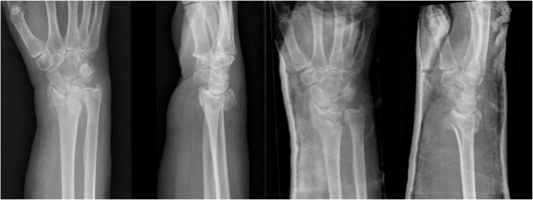
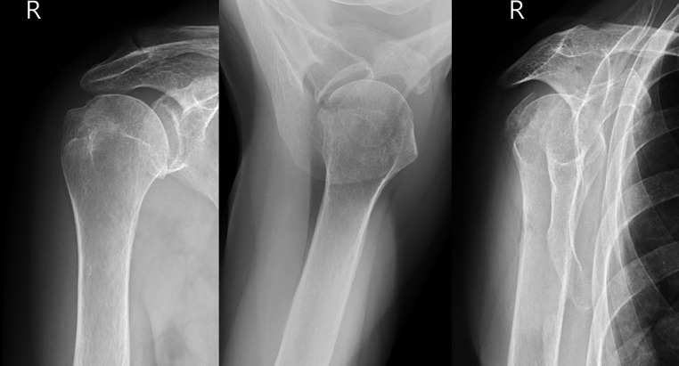
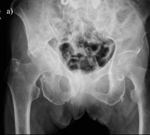
Source note: the radiographs are cropped from open-access clinical articles with source details collected in the radiograph library.
The big idea
After a fracture, it is natural to focus on the bone that broke. That matters. But if the fracture happened from a simple fall or low-energy event, there may be a second question: did the bone break because the skeleton had less reserve than expected?
Fracture liaison service research exists because this second step is often missed. High-priority fracture references describe a handoff problem: the broken bone is treated, but osteoporosis evaluation, fall-risk work, medication review, and secondary prevention may not happen unless the system connects the dots.1,2,10,11
Two tracks should run at the same time
Visual explainer: the fracture can be the doorway
A fall from standing height, a wrist fracture, a shoulder fracture, a hip fracture, or a spine compression fracture.
The broken bone may reveal low bone density, lower bone quality, fall risk, medication effects, or another medical contributor.
Use the fracture as a reason to ask better questions before another fracture happens.
What may be part of the workup
Measures bone density at clinically important sites and provides T-scores/Z-scores. It is a starting point, not the whole story.5
Sometimes used to look for vertebral compression fractures, which can change risk assessment.
Vitamin D, calcium metabolism, kidney function, thyroid/parathyroid issues, blood counts, and other tests may be considered by a clinician.
Vision, balance, medications, footwear, home hazards, strength, and confidence all matter for preventing fractures.
Common questions
Does every fracture mean osteoporosis?
No. A fracture does not automatically mean osteoporosis, but some fractures should trigger a bone-health conversation, especially after age 50 or after a low-energy fall.
Should I wait until the fracture heals?
Fracture healing and bone-health evaluation can often proceed in parallel. Timing depends on the injury, surgery, mobility, and your clinical team.
What if my DXA does not say osteoporosis?
Many fractures occur in people whose T-score is not -2.5 or lower. Age, prior fracture, falls, medications, and FRAX-style risk assessment can change the conversation.8,9
Who should own the follow-up?
That varies. A fracture liaison service is designed for exactly this handoff, but primary care, endocrinology, rheumatology, orthopedics, or another clinician may coordinate.
Printable tools
References
- Napoli N, Chandran M, Pierroz DD, Abrahamsen B, Schwartz AV, Ferrari SL. Coordinating multidisciplinary care: improving outcomes after fragility fractures. N Engl J Med. 2025;392(4):379-391. doi:10.1056/NEJMra2304245.
- Napoli N, Chandran M, Pierroz DD, Abrahamsen B, Schwartz AV, Ferrari SL. Coordinating multidisciplinary care: improving outcomes after fragility fractures. N Engl J Med. 2025;392(4):379-391. doi:10.1056/NEJMra2304245.
- Matzkin EG, DeMaio M, Charles JF, Franklin CC. Diagnosis and treatment of osteoporosis: what orthopaedic surgeons need to know. J Am Acad Orthop Surg. 2019;27(20):e902-e912. doi:10.5435/JAAOS-D-18-00600.
- Morin SN, Leslie WD, Schousboe JT. Osteoporosis: a review. JAMA. 2025. doi:10.1001/jama.2025.6003.
- El Maghraoui A, Roux C. DXA scanning in clinical practice. QJM. 2008;101(8):605-617. doi:10.1093/qjmed/hcn022.
- El Maghraoui A, Roux C. DXA scanning in clinical practice. QJM. 2008;101(8):605-617. doi:10.1093/qjmed/hcn022.
- Kanis JA, Johansson H, Harvey NC, McCloskey EV. A brief history of FRAX. Arch Osteoporos. 2018;13(1):118. doi:10.1007/s11657-018-0510-0.
- Bawa HS, Weick J, Dirschl DR. Anti-osteoporotic therapy after fragility fracture lowers rate of subsequent fracture: analysis of a large population sample. J Bone Joint Surg Am. 2015;97(19):1555-1562. doi:10.2106/JBJS.N.01275.
- Napoli N, Chandran M, Pierroz DD, Abrahamsen B, Schwartz AV, Ferrari SL. Coordinating multidisciplinary care: improving outcomes after fragility fractures. N Engl J Med. 2025;392(4):379-391. doi:10.1056/NEJMra2304245.
Educational Use Only
This website is educational. It is not a medical practice, telemedicine service, or a substitute for care from your own clinician.